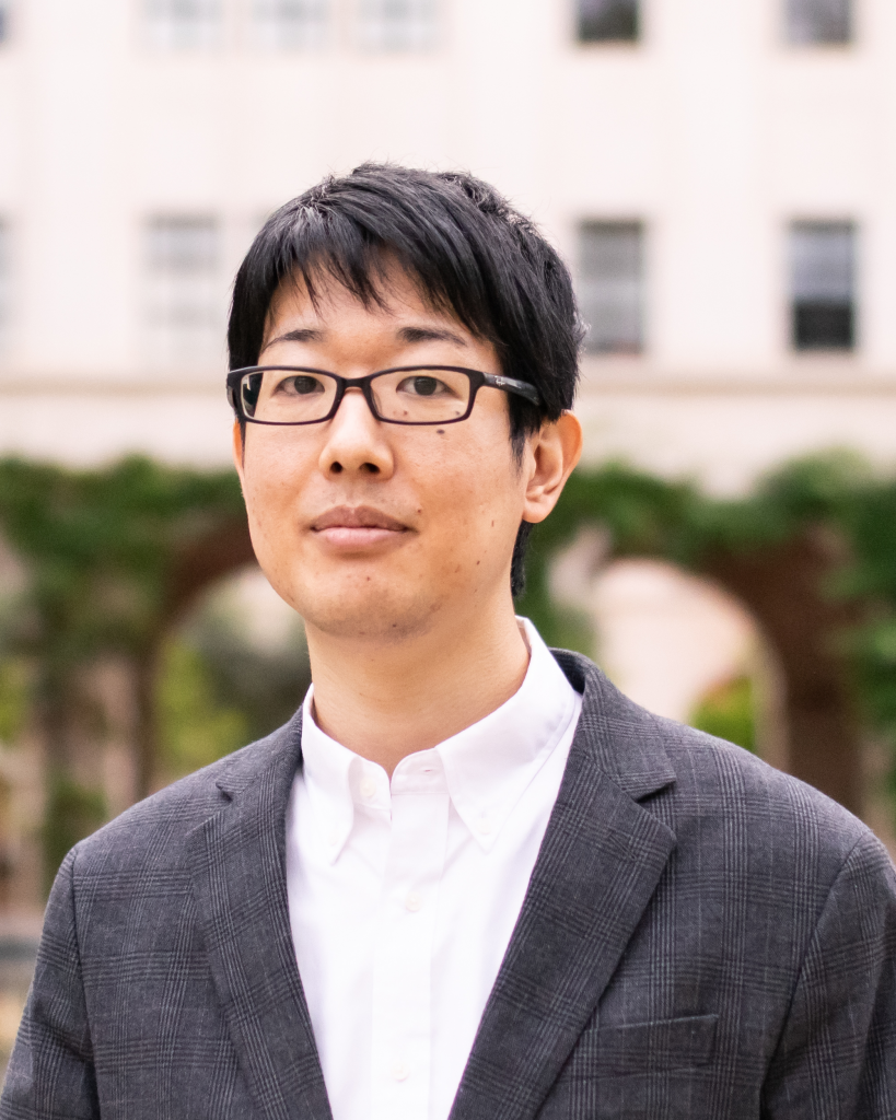
Yodai Takei
Division of Biology and Biological Engineering, California Institute of Technology, Pasadena, USA
Essay
Imaging nuclear architecture in single cells: Multiplexed imaging technologies uncover precise 3D maps of single nuclei
Abstract
Various molecules such as DNA, RNA, and proteins are spatially organized within cells
in our body. Inside each cell, the three-dimensional (3D) structure of the nucleus is
particularly vital for comprehending different cell types and states. Nevertheless,
our understanding of 3D nuclear organization has been limited by the lack of high-
resolution spatial multiomics technologies. To overcome this, I have developed
multiplexed imaging-based approaches, allowing transcriptome- and genome-scale
imaging of individual molecules within single nuclei.
Utilizing these sequential fluorescence in situ hybridization (seqFISH) methods, I
obtained precise 3D maps of the nucleus, incorporating nascent transcriptome, genome
structures, and subnuclear structures in various cell types. Collectively, my thesis work
establishes a crucial foundation for understanding diverse cell types and states through
the principles of nuclear organization.
Biography
Yodai grew up in Yamanashi, Japan, and received his bachelor’s and master’s degrees
in Pharmaceutical Sciences from The University of Tokyo. He earned his PhD in
Biology from California Institute of Technology. He worked with Long Cai to develop
imaging-based single-cell multi-omics technologies. Using these new technologies, he
investigated the 3D organization of the nucleus in diverse cell types, revealing nuclear
features emerging from the interplay of DNA, RNA, and protein molecules.
Yodai is currently a postdoctoral scholar in Michael Elowitz’s lab at California Institute
of Technology, studying spatiotemporal and combinatorial aspects of chromatin
organization and gene regulation. Outside of the lab, he enjoys reading, running, and
traveling.
